In a groundbreaking study, scientists have observed brain cells forming groups and working together, akin to friendships, using advanced microscopy techniques.
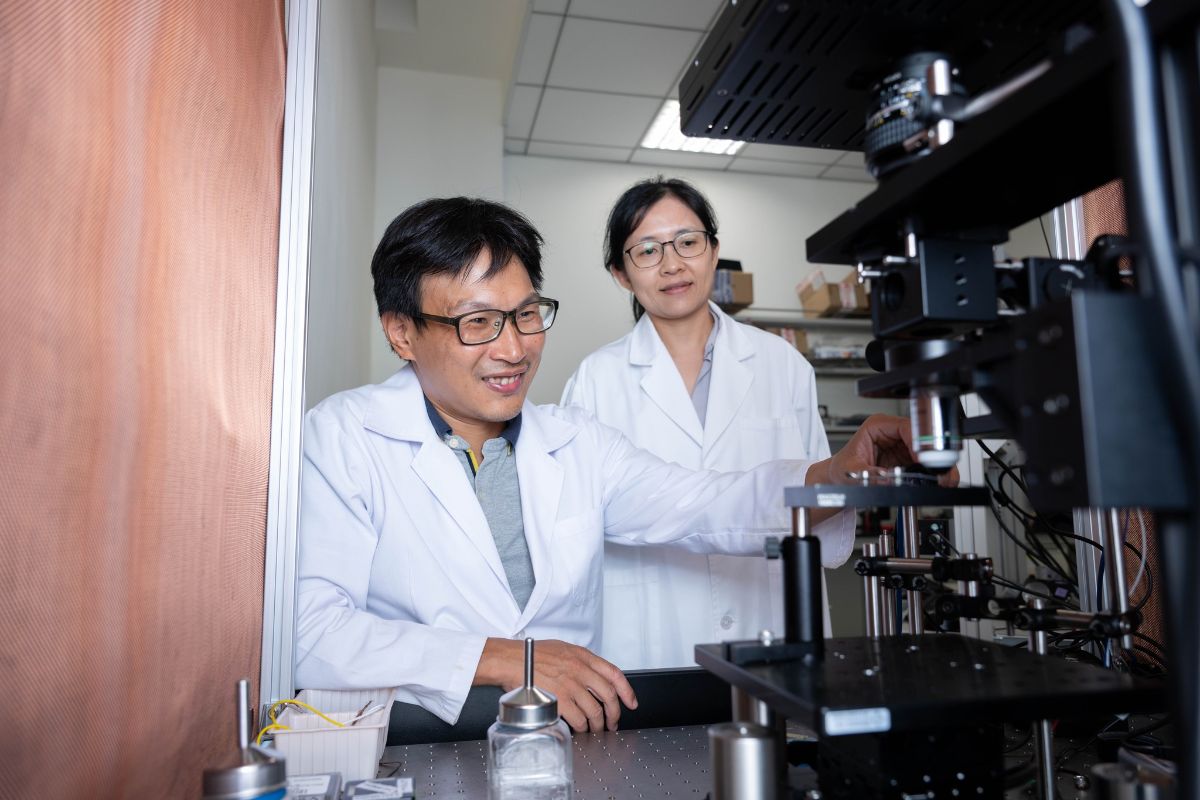
Published in this month’s issue of Neuron, the study from the Institute of Neuroscience at National Yang Ming Chiao Tung University (NYCU) reveals how neurons exhibit collective behavior. Assistant Professors Tsai-Wen Chen and Bei-Jung Lin led the research team that discovered neurons activating synchronously, indicating a preference for specific cells to ‘team up’ during activation.
Rare Interneurons Unveil Group Dynamics, Facilitating Brainwave Formation
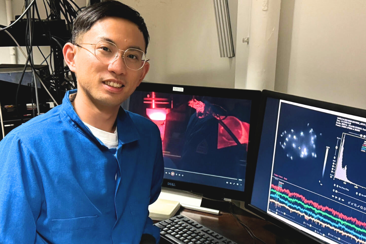
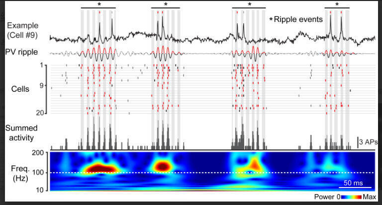
Interneurons, rare and previously studied through sporadic electrical signals from implanted electrodes, were likened to “finding a needle in a haystack,” according to Bei-Jung Lin. “Recording even a single cell could take a month, making interaction studies challenging.” The team used voltage imaging with fluorescent proteins to record up to 26 interneurons, unveiling their interaction patterns.
The study found that interneurons do not activate randomly but tend to fire together, suggesting they find ‘like-minded friends’ to transmit electrical signals. “Much like an orchestra following a conductor,” Tsai-Wen Chen explained, “interneurons are crucial for inhibitory neurotransmission in the brain and play a key role in brainwave formation.” Brainwaves arise from synchronized electrical activity across numerous neurons, detectable from the scalp. Interestingly, even without reaching the activation threshold, neurons displayed group activity under the microscope.
Innovative Imaging Technology Sheds Light on Neural Activity and Brain Function
To overcome the challenge of directly observing voltage with a microscope, the research team led by Tsai-Wen Chen and Bei-Jung Lin collaborated internationally to develop voltage-sensitive fluorescent proteins. These proteins, delivered to neurons using adenoviruses as carriers, allow the cells to glow upon activation.
Additionally, to capture the highly brief neural impulses, the research team designed and set up an ultra-high-speed imaging system capable of capturing 2,000 frames per second. These technologies and facilities are now established at NYCU.
Brainwaves are critical signals in perception and memory functions. This new technology allows scientists to observe collective neural operations in living animals, revealing the intricate coordination essential for understanding brain functionality.